There is a disorder (it
may actually be two or three) in the canine similar to the “wobbles” in
thoroughbred horses. Much has been written on this, with various
descriptions such as cervical vertebral instability (CVI), cervical
spondylopathy, stenosis of the cervical vertebral canal, caudal cervical
spondylomyelopathy, and even the mouthful, caudal cervical vertebral
malformation/malarticulation. It was once referred to (incorrectly,
perhaps?) as spondylolisthesis. However, no term is more used, more
succinct, more descriptive, or even perhaps more accurate, than “wobbler
syndrome”. In the horse, about 12% of this syndrome can be blamed on
osteochondrosis and here, too, there may be some parallels with the dog.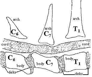
Cervical Vertebral Instability (CVI or “Wobbler Syndrome”)
That we may be dealing with two very similar disorders or two variations of a disorder is indicated by the differences seen in early studies on middle-aged and older Doberman Pinschers, Basset Hounds, and later work with Great Danes. The earlier work pointed at instability, subluxation of the vertebrae, and a tendency for one of the vertebral bodies (the actual bone segments of the spine, not including the material between) to lose bone, ride up onto the one in front of it, and thus compress the spinal cord from the bottom. See the figure provided. This would be a little like a car with an angled bumper running into another car ahead of it, its front bumper sliding up and over the rear bumper of the other vehicle and smashing into its trunk and taillights, although in slow motion. Mostly large breeds have been involved such as those named above, plus Saints, Old English Sheepdogs, and Rhodesian Ridgebacks, though infrequently in smaller breeds, also.
Another abnormality is seen in the spinal column of dogs with Wobbler Syndrome: a partial closing or narrowing of the canal by abnormal thickening of the vertebral body surrounding it. Stenosis is a squeezing or partial closing of a tubular structure such as an artery, heart arch, or, as in this context, the spinal canal (where the spinal cord runs through the vertebrae). It is congenital (found at birth), developmental (gets worse), and degenerative (has destructive effects) in man and probably so in dogs as well. Stenosis has been seen in the cervical and lumbar vertebral canal and the intervertebral foramina (spaces between the vertebrae), and may be “silent” not giving rise to complaint, in many individuals unless accompanied by other factors such as protrusion of a disc, spinal instability, or movement such as the extreme flexion or extension of the neck or other part of the spine.
Most of the lesions in early studies were between the sixth and seventh cervical (neck) vertebrae, which are designated C6 and C7. It has been thought that there was an inherited malformation of these spinal segments (vertebrae) with possibly a simple recessive trait but more recent evidence indicates more genes are involved in somewhat more complex ways. We know a little more about osteochondrosis and the etiology of osteophyte formation now.
Clinical Signs
At the point of greatest cord compression, the damaged ascending sensory neurons (those nerve cell carrying electrical impulses to the brain) begin to die. Their myelin sheaths deteriorate, and confusing impulses cross over from one axon to another, in effect making the brain and cord “think” they are coming from someplace else, and thus the return messages to contract certain muscle fibers may be sent to some of the wrong places. This gives rise to much of the missteps and poor movement. Many of the axons (main conductors) also die, and loss of sensation results.
Simultaneously, descending motor neurons are affected the same way, so fewer of them function from that part back to the muscles. Therefore, the dog may seem not to know where its limbs are, drags its toes in a manner similar to those with GSD myelopathy or stands on the top surface of one of its rear paws, has poor coordination especially in the pelvic limbs, may stand wide and, if the thoracic limbs are affected, may have a prancing gait. In severe cases, the dog falls down easily and has a hard time getting up on its feet. The syndrome affects both sides of the body equally.
The next figure represents a neuron (nerve cell), which can be very long as in some peripheral nerves or very short as in some brain cells. Most have myelin “insulation” covering the axons that conduct electrochemical messages much the way electrical wires conduct electricity in your house or appliances. Sensory nerves have different endings than do motor nerves, and one sensory ending may vary from another way.
The descriptive terms “ataxia” and “spastic paresis” in this disorder refer to weakness and partial paralysis with incoordinate motion, and these are seen principally in the rear. When cervical spinal cord damage is further toward the head, the forelimbs and even perhaps the diaphragm may be involved, but in canine Wobblers, the lesion is usually found in the caudal (rearmost) cervical vertebral spaces such as C6-7, rarely C5-6, although in Basset Hounds the same or a similar syndrome is associated with the cord pinching occurring around C2-3. Wherever it exists, it may cause an abducted (limbs move away from each other) and sometimes fast-beat gait with stumbling especially when turning. The ataxia, of course, is due to the damage to the ascending neurons and the jerky movement and paresis to damage to the descending neurons, both at the point of cord compression.
Cause
It was discovered very early in man and described in the dog by 1967 that vertebral canal stenosis is a cause of spinal cord compression and these researchers considered deformation of the vertebral bodies to be the cause of that stenosis. Others have looked at this and similar problems in a variety of breeds.
About the same time as the above work and a little later, another cause of spinal canal stenosis was proposed: a deformation of the vertebral arches (that part of the segments covering the canal), as well as the discs, the processes, and articulations in the joints between the bone segments. Other things happening at the same time and possibly contributing to stenosis or associated with it otherwise include hypertrophy (overgrowth or thickening) of the flavum ligament or of the dorsal longitudinal ligament or of the dorsal annulus. Simple poor alignment and malarticulation have also been blamed or implicated. Another cause of compression of the cord is the CVI (cervical vertebral instability) mentioned earlier, and identified by various names such as spondylolisthesis and vertebral subluxation.
In the earlier work on CVI, instability was the diagnosis when greater flexion between two vertebrae than “normal” was evident. When we speak of flexion, we mean the bending of joints so that the limb or extremity is “folded” toward the centerline of the torso, while extension is a straightening-out away from the rest of the body. In speaking of the neck, flexion is the bending of the head downward toward the sternum, and extension is the bending up as if stretching to reach over the back. How valid is the diagnosis of neck instability as “shown” in flexion, was brought into question in 1977 by Wright who found the abnormal angulation (one bone starting to slip or ride up on another) in many dogs who had no clinical signs of cervical spinal problems. It had been standard practice to bend the neck down fully and see if there were any irregularities or subluxation on the radiograph. These pictures were compared to the neck in a neutral position (same as it would be carried in standing). These pictures were almost invariably taken in a lateral view (from the “side”; with the dog in lateral recumbency), although some people showed how useful a ventrodorsal view could be in demonstrating lateral compression of the cord.
In 1982 Olsson, Stavenborn, and Hoppe in Sweden studied Great Danes and found that the ones with wobbler syndrome had radiographically demonstrable lesions only when the neck was in extension, which ran counter to the experiences previously reported. They did not find any CVI or vertebral body deformation, except for a slight deformation in the vertebrae of one dog, and guessed at a number of possible reasons. Their work was only with Great Danes, and previous studies had also included Bassets and Dobes, so perhaps there is a different genetic pathway for some breeds than for others. The syndrome in Danes occurs mostly in young dogs and in Dobes they occur mainly in older dogs. The 1982 Swedish study involved “wobbler” dogs from 4½ to 24 months. Both plain radiographs and some made after the injection of a dye for myelography were studied, with the necks in all positions. The euthanized dogs’ spinal systems were then studied for comparison.
Even normal Danes have relatively smaller ventrodorsal height to the spinal canal, and larger prominent intervertebral joints compared to many other breeds. When they looked at a dog without wobbler signs and increased C3-4 flexion as seen on a regular film, the myelogram didn’t indicate any pinching of the cord, even though it looked as if the bone could have done so. The wobbler dogs with increased flexion between two vertebrae showed no pinching, either. The picture that came out of this work in Sweden is that compression of the cord is most severe when the neck is extended. It also appeared that the compression and stenosis took place at the cranial end of the vertebral canal, where the height was less and the “roof” was pushed down onto the cord when the neck was lifted (extended). This is probably why many wobblers will hold their heads down, by the way, but that is not the best diagnostic sign. Often, Danes with wobbler syndrome have an abnormal vertebral arch (between the “neckbones ”) that is plump and longer, and frequently it is seen in association with deformed and asymmetrical articular processes. Many also have considerable osteophytic deposits that may contribute to further compression. Compression was more dynamic than static, meaning other forces combined with the malformation to produce the compression, such as disc protrusion and even the normal movement of the head and neck. Possibly the most important finding of this research, beyond the hint of breed and genetic differences, is that cord pinching might not be demonstrated in wobblers unless myelography is used in conjunction with radiography and pictures are taken with the neck in different positions.
The breeder/owner can make a tentative diagnosis based on symptoms before taking the dog in for myelograms. But you want to remember that there are other problems, and you should differentiate between them. If the dog has no pain, but the unstable gait described, it is very possibly wobbler syndrome. If pain and hypersensitivity are present, your dog may instead have cervical disc protrusion syndrome. The pain probably comes more from the secondary inflammation that results when the disc’s nucleus pulposus tissue extrudes into the epidural space in the canal and calcifies with this “hardened cement-like” material acting much the way osteophytes do in irritating and abrading the surrounding soft tissues. In the wobbler, inflammation is not much of a problem if at all.
In the important 1974 work at Cornell on Great Danes, joint problems, and nutrition, evidence indicated that vertebral body deformation is a manifestation of osteochondrosis brought on by rapid growth and overnutrition. Olsson and colleagues commented that some of the changes seen in the cartilage between the vertebrae have similarities to osteochondritic changes seen in other joints, and implies that high-energy, high-calorie “rich” diets may, in certain dogs and breeds genetically predisposed to these disorders, bring on the osteochondrosis responsible for the stenotic myelopathy (pinching and disease of the spinal cord) seen in animals.
Other environmental factors may worsen a congenital or hereditary problem. It has been theorized that the very heavy head of a Thoroughbred horse, a Great Dane, or a Basset Hound put great stress on certain vertebrae during early growth, but there are too many questions to give much credence to that. Dobes certainly don’t have heavy heads, and different vertebrae are involved in different breeds. Separate genes and locations may be involved in Bassets compared to other breeds. A cooperative pedigree and clinical study program between breeders and some veterinary school teams could provide more accuracy in detection and improve some breeds of dogs through prevention of disease.
Treatment of Wobbler Syndrome
Earlier treatments have included fusion of adjacent vertebrae with bone grafts and bone cements, or simply the use of anti-inflammatory drugs, but improvements were needed. A technique developed at the U. of TN in 1983 calls for drilling a slot between the two segments and filling the gap with about ½ to 1-inch of bone cut from the pelvis. What makes it different is the use of stainless alloy rods with hooked ends attached to other vertebrae, and a threaded section with nuts that can be tightened to adjust tension. About 75% of the dogs treated this way at the U. of Florida vet school regain at least partial use of their legs, a much better prognosis than has previously been had. Fewer dogs now face euthanasia.
Fenestration is an operation in which a hole or channel is cut (usually in a disc) so that the spinal cord or large nerve at the ganglia has more room. But unless something is done to prevent continued or repeated cord compression, it is rarely successful. If the two bones that squeeze the nerve are spread apart (distracted) and the space and relationship of the bones stabilized, there is much greater chance of success. A technique developed at Tufts involves a plug of polymethyl methacrylate — in effect, a wad of “super glue”. Anchor holes are drilled in opposing faces of adjacent vertebrae where most of the disc has been cut out (but some left to bridge and cushion the bones). After the glue plug hardens, the distraction device is removed, and some cancellous bone (harvested from the greater tubercles of one or each humerus) is grafted onto the bottom, causing what would appear to be a man-made spondylosis (bridge) on the ventral surfaces. Of course, there is no flexibility between these two “fused” segments, but no misalignment and consequent repeat of the cord compression, either.