Elbow Disease In Growing Dogs
by Roger Lavelle |
|
© Copyright May, 2000 - 2005 |
INTRODUCTION
Elbow disease is the preferred term to be used when talking about elbow problems in growing dogs. Unfortunately "elbow displaysia" was the name given to the condition of ununited anconeal process and this term is closely linked in this way in the minds of most veterinarians and some dog breeders.
Elbow disease is a general term to denote joint problems in growing dogs and it includes ununited anconeal process (UAP), fragmented medial coronoid process (FCP) and osteochondrosis of the medial condyle of the humerus (OCD). These are the three most important conditions although there are a number of uncommon conditions included in the term.
Elbow disease has received increasing publicity in recent years due to the high prevalence of foreleg lameness localised to the elbow joint and the realisation that elbow disease has a hereditary basis. There are two important situations to discuss, firstly the management of clinical elbow disease and perhaps more importantly, the monitoring of elbow disease by breed clubs.
1. CLINICAL FEATURES
Elbow disease is a problem of growing dogs and the clinical signs of the three main problems are somewhat similar. The earliest problem recognised was UAP. The German Shepherd Dog and the Basset Hound are the two main breeds involved, although any middle size or larger breed may be affected.
The anconeal process sometimes grows as a separate ossification centre and it is usually recognised as such at around 70 days. It is usually united to the main part of the ulna by about 140 days. However, dogs which have UAP do not necessarily show lameness. Thus dogs with elbow lameness and an UAP which are older than 140 days would be considered to be exhibiting signs relating to this condition. the radiological diagnosis is straightforward and there may be osteoarthritic change in addition to the presence of UAP. It is also possible that dogs with UAP could in addition have FCP and/or OCD.
Dogs with OCD or FCP may present with lameness earlier than UAP cases. there have been cases as young as 3-4 months, but the 5-8 months category would be more common. Cases continue to be presented up to 18 months or older, but dogs of some years presumably present with exacerbation of underlying osteoarthritis.
The forelimb lameness may be unilateral or bilateral. There is often pain on manipulation of the elbow and a reduction in range of movement. There may be swelling of the elbow joint. The specific diagnosis can sometimes be made on x-ray examination, but more frequently the diagnosis relies on the presence of osteoarthrosis of the elbow joining which is the result of either a primary FCP and/or OCD. A variety of views can be used to examine the elbow joint and they may pick up the primary problem. It is more likely that OCD will be identified rather than FCP. Despite high quality x-rays, it is impossible to identify all FCP and OCD lesions without using special techniques and the diagnosis is based on the clinical findings and the radiographic changes of osteoarthrosis.
2. MANAGEMENT OF CASES OF ELBOW DISEASE
Ununited anconeal process can be managed by either removing the UAP or attaching it firmly to the ulna using a lag screw. The former is the simplest and normally gives excellent results. Fragmented coronoid process and OCD lesions may produce a temporary lameness that responds to rest or medical treatment. If the lameness persists then surgery is indicated. This involves a medial approach to the elbow joint on the opposite side to surgery for UAP, and both the medial coronoid process of the ulna and the medial condyle of the humerus must be examined carefully. One or both lesions may be present and they can cause "kissing lesions" on the opposite side of the joint. The surgical results vary with breed and age, but many dogs will settle satisfactorily.
3. MONITORING OF ELBOW DISEASE
The monitoring of elbow disease has been undertaken for some years now in some parts of the world, particularly on the Continent of Europe. The breeds which have serious problems with hip displaysia appear to have elbow disease as an equally important problem. There have been a number of papers suggesting the UAP has an inherited basis, but little has been done to control it. It was only when studies on elbow arthrosis indicated that elbow disease was inherited that action began to be taken to monitor its incidence.
Early work documented the presence of elbow arthrosis, the secondary osteoarthritic change, and various workers graded the degree of change similarly to what was happening with hip displaysia. The increasing international awareness of the importance of elbow lameness in growing dogs led to the formation of the International Elbow Working Group. The aims of this group were to establish an internationally accepted radiological interpretation system and to encourage research into the cause(s) of the development of the primary problems. The working group is independent but has held its meetings in conjunction with the World small animal Veterinary Association conferences. It is growing in membership and recently played the major role at the meeting organised by the WSAVA and reported in the VCA gazette by Dr. Robert Zammit, the ANKC representative at the meeting.
The International Elbow working Group's guidelines for monitoring of elbow disease were documented in the Gazette. there is simply a requirement for good quality flexed lateral elbow views which are assessed for the presence of arthrosis. This view will readily identify UAP but only rarely picks up FCP and OCD. There is not really a problem as at present no other causes of disease have been commonly identified. However there is a problem in the reporting of findings where there is no definite differentiation between UAP cases and those with degenerative joint disease. As mentioned earlier, it is possible to find UAP in the presence of other primary elbow problems.
It is all very well to monitor the elbow disease but unless some constraints are put on breeding, then there will be a lot of x-rays taken but no improvement in the prevention of lameness in the breeds affected. The research from the continents of Europe, Britain, Australia, and USA has shown that elbow disease is inherited . There is also information to show that those dogs with the more severe lesions are most likely to produce puppies with serious elbow disease. Consequently grade 3 elbow disease dogs should not be used for breeding and the grade 2 cases should be considered as serious risks.
The suggested age for x-ray examination is 12 months when the hip x-rays are taken. It is likely that the severity of the osteoarthritis will increase with age and consequently for the monitoring programmes at present, dogs should be examined when young. It may be that later the age for screening will be raised, but this will mean an alteration to the current breeding programmes. Owners and breeders need to be aware that not all breeds behave in the same way in regard to elbow disease. for example, most published reports suggest that surgery in Rottweilers has little benefit compared to medical therapy, whereas our results in the Labrador Retriever have been rewarding. Certainly elbow disease is not as straightforward a problem to handle as OCD of the shoulder and in some cases, the severity of the chronic elbow disease may lead to dogs being destroyed.
Responsible owners and breeders of dogs of the breeds where elbow disease is a recognised problem should consider monitoring the elbows in the same way as they monitor hip displaysia and eye disease. The x-ray examination is simple and the Australian Veterinary Association will shortly have application forms for elbow disease assessment. The German shepherd dog Club has forms available to enable assessment of both hip and elbow x-rays or either alone through the club schemes.
 Nil
Arthrosis (Grade 0)
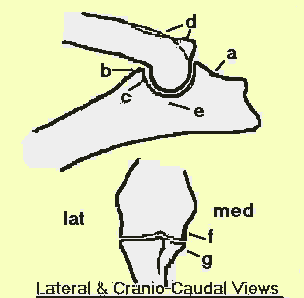
Minimal Arthosis (Grade 1) = one or more of
the following findings:
(a) less than 2 mm high osteophyte formation seen on the dorsal edge of
the anconeal process (b) minimal osteophyte formation (less than 2 mm in
any direction) on the dorsal proximal edge of the radius (c) or the
dorsal edge of the coronoid process, (d) or the leteral palmar part of
the humeral trochiea; (e) sclerosis in the area caudal to the distal end
of the ulnar trochlear notch and to the proximal
Moderate Arthosis (Grade 2) = one or more of
the following findings:
(a) osteophytes 2 - 5 mm high on the anconeal process (b) moderate
osteophyte formation (2 - 5 mm in any direction) on locations b, c, d.
Severe Arthosis (Grade 3) = one or more of the
following findings:
(a) more than 5 mm high osteophyte formation on the anconeal process (b)
severe osteophyte formation (more than 5 mm in any direction) on
lcations b, c, d.
Additionally - in cranio-caudal radiographs osteophytes are most easily seen on the distal, medial part of the humeral condyle (f) and the medial part of the coronoid process (g).
University of Melbourne