considered to be the hip assessment method that will bring a genuine improvement in hips
PennHIP Radiographic Evaluation at a Glance
The PennHIP method is a different way to assess, measure and interpret hip joint laxity. It consists of three separate radiographs: the distraction view, the compression view and the hip-extended view (see below). The distraction view and compression view, developed by Dr. Smith, are used to obtain accurate and precise measurements of joint laxity and congruity. The hip-extended view is used to obtain supplementary information regarding the existence of DJD in the hip joint.
|
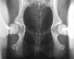
Distraction View |
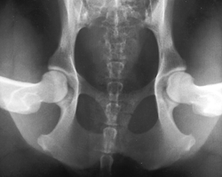
Compression View |
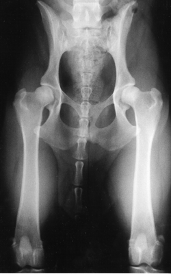
Hip-extended View |
|
|
|
|
|
The radiographs above and to the right are of the same dog, yet the hip joint laxities (looseness) in each view look very different. Notice that the hips in the distraction view (top left) appear to be much looser than they do in the hip-extended view (right). On average the distraction view has been shown to reveal 2.5 - 11 times more hip laxity (depending on breed) than the hip-extended view. Also the PennHIP method can measure the laxity of a hip joint with greater precision than the hip-extended method. The degree of hip joint laxity, as measured by the PennHIP method, has been shown to be the most important risk factor in determining whether a dog is prone to developing CHD.
VISIT
THE PenHIP WEBSITE |
||
Dr. Gail Smith Interviewed about PennHIP
Dr. Smith was interviewed by Lisa Stahr, host of Special Pets, Special Needs. Gail talks about the PennHIP method and how it can predict the likelihood of getting Degenerative Joint Disease/Osteoarthritis later in life. The subjects of heredity and selective breeding for better hips were also discussed. This interview was geared toward the layperson and explains the concepts of hip dysplasia and breeding in understandable day-to-day language,LISTEN TO THE INTERVIEW "HERE"